News

Making An Effort

When I first started this website, I had intended it to be little more than a hub for my photography. My images were posted to a generic page and poorly organized, my news posts were little more than ramblings and information related to sales and new images I had made, and I honestly had little to no idea what I was really wanting from a website.

Over the past two (three?) years, my abilities as a photographer and artist, as well as a scientist, have noticeably grown – if not to anyone else, then certainly to myself. I’ve taught myself new imaging techniques, I’ve picked up illustrating (albeit not quite good at it but making the effort to improve), I’ve made new contacts, I’ve began carving out a more solid niche in my own scientific community, and – perhaps most importantly – I’ve became more organized.

This website has developed in many ways since its inception back in 2016. Pages are organized, my images are neatly displayed (at least I think they are) by category and again in the order of newest in front, oldest in back, my sketches are organized by year and day of creation rather than sloppily posted in some haphazard fashion, I actually update my C.V. at least once a month, and information regarding me as a scientist can actually be found on the site now. Hell, I’ve even made a couple of figures for my own benefit that I’ve hosted here in hopes others may also benefit from them.

Point being, this site has changed a good deal, and I along with it. Something that I sincerely look forward to saying again in three years from now. I feel that it be only appropriate if I also begin using my News page for relevant … news. From presentations that I’ll be giving, to future publications, to awards when I receive them – I’ll be making my best effort to actually post information regarding me as a professional – be that as a scientist or an artist.

Thank you all for your support over the past few years and here is to another year of growing and making efforts to better improve ourselves.

-M. Chaise Gilbert


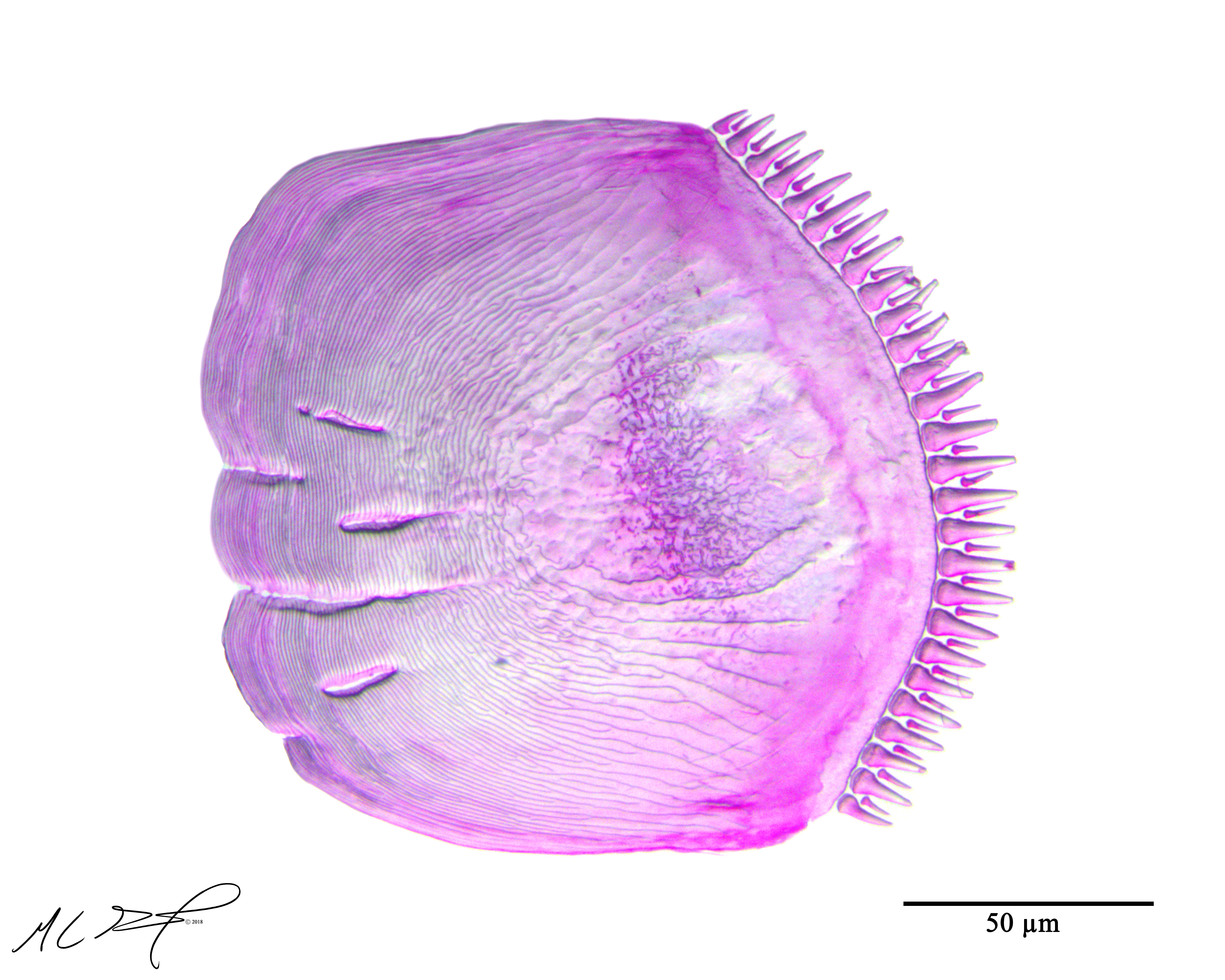
Ctenoid Scale from Pseudanthias squamipinnis

I found this ctenoid scale by accident and I couldn’t bring myself to simply rinse it down the drain or dispose of it. So, I imaged it as best I could and put it up on here for both my own, and everyone else’s, gratification. Enjoy!

Full Figure

Cheek Scale from <em>Pseudanthias squamipinnis</em>, a sea goldie. MC Gilbert, 2018.
Preview of a ctenoid scale taken from the cheek (just post-orbital) of Pseudanthias squamipinnis. Specimen was cleared and stained prior to scale removal.

Palatine teeth on the stripetail darter

I made a figure to help me distinguish the difference between vomerine teeth and other teeth on the roof of the mouth in darters. I hope this is helpful to others as well.

Image Page

Palatine Teeth
Preview of pterygoid and vomerine teeth in Etheostoma kennicotti, the stripetail darter. A: ventral, anterior view. Window draws attention to the location of teeth along the pterygoid process and vomer (on the roof of the mouth). B: view of palatine teeth, from ventral view. Teeth on left pterygoid process marked by the left arrow; vomerine teeth marked by the right arrow. Scales bar represents 500 µm. C: right lateral side, anterior. Window draws attention to location of palatine teeth. Scale bar represents 2 mm. D: palatine teeth at increased magnification. Teeth on left pterygoid process marked by the posterior arrow; vomerine teeth marked by the anterior arrow. Scale bar represents 500 µm.Big thanks to D Navon, D Jockel, B Richardson, RC Albertson, and S Huskey for comments and feedback. 

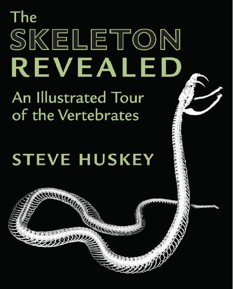
“The Skeleton Revealed” by Steve Huskey

I am excited to announce, and help spread the word, that Dr. Steve Huskey’s new book, “The Skeleton Revealed”, is published and now available for purchase! I had the pleasure of working with Steve when he served as one of my committee members and as a mentor during my time as a graduate student at Western Kentucky University. Steve provided me with ample opportunities to further my understanding of vertebrate anatomy, in addition to providing me with many of the specimens that you now see up on this website.

Steve’s book can be purchased through John Hopkin’s University Press, as well as on Amazon (links take you directly to the book).  For a sneak peek into the amazing skeletal displays presented in his work, visit “The Skeleton Revealed” @ Issuu.com .

cover