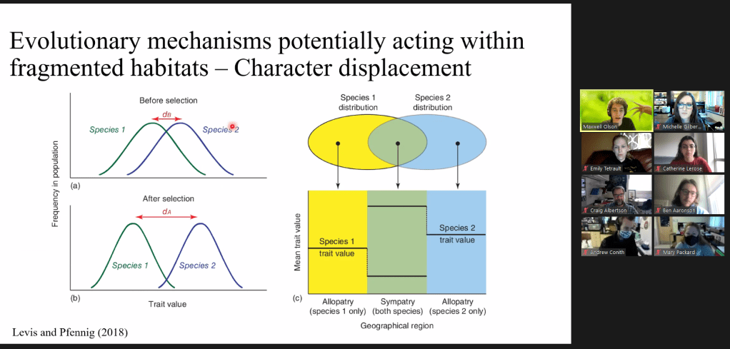
Mr. Max (Maxwell) Olsen just successfully defended his undergraduate honors thesis! Max has worked with me for the past two years in the Albertson lab, primarily aiming to quantify shape in the endangered, and highly fragmented, populations of Pecos gambusia (Gambusia pecosensis).
From being a hands on undergraduate teaching assistant, to a dedicated and hypothesis driven researcher, Max has been an outstanding individual to work with. Aside from now preparing our findings for publication in a peer-reviewed journal, he is preparing to start the next chapter of his life as he pursues his doctoral studies at Clark University. There, he will explore the morphology and evolution of limb loss across vertebrate taxa.
Ms. Catherine Lerose, an undergraduate honors student that has been working with me in the Albertson Craniofacial Lab for the past two years, has successfully defended her honors thesis! Catherine, who has worked on a number of projects since first joining the lab, took interest in the bramid project we had been developing.
Catherine has been an outstanding student to work with and watch grow over the past years. Her contributions have earned her a place as a co-author on a recent paper, as well as on an upcoming manuscript (stay tuned!).
Catherine is currently looking to expand her knowledge of evolutionary theory, but also take a dive into ecology and behavior, by applying to graduate student positions.
Best of luck, Catherine, and remember that we are here for you in your journey!
Ms. Catherine Lerose proudly holding a bramid (Brama sp japonica) specimen while visiting the Harvard Museum of Comparative Zoology.
So much has happened over the past several months that I’ve forgotten to update this page!
Nevertheless, I’ve great news to share. Since July 2020, I’ve had three papers accepted for publication!
The first, titled “Rapid morphological change in multiple cichlid ecotypes following the damming of a major clearwater river in Brazil”, uses geometric morphometrics (a fancy way of using math to study how geometric shapes differ across organisms) to see how cichlid populations have changed over the past 50 years. As the titles states, dams are involved. One in particular, and a major predicted driver in our study, is quite large, and, in the area it exists, has changed the Tocantins river from being shallow and clear, to being a massive reservoir. In short, we find that all five cichlid species in our study have changed shape over the past 50 years! For more detail, check out the full paper here!
Our second, titled “Extreme Morphology, Functional Trade-Offs, and Evolutionary Dynamics in a Clade of Open-Ocean Fishes (Perciformes: Bramidae)” takes advantage of a small, but rare family of fishes that have some unique, but clear family relations. One of the groups, the Ptericlinae, have exaggerated medial fins that we feel can provide unique insights into how some traits are prevented from changing during evolution based on the history of the family. In short, we think history matter and we explore and explain why, here!
Figure 8. Reconstruction from µCT scans of a representative Pterycombus petersii, standard length 7.9cm
Our third, titled “Ciliary rootlet coiled-coil 2 (crocc2) underlies evolutionary divergence and plasticity of cichlid jaw shape”, uses zebrafish to investigate a possible mechanism plasticity (the ability for an organism to change in response to a unique environment). We find and discuss a molecular mechanism and present our case for why we think it may play a role in plasticity. For more, check out our paper here!
Figure 1. Functional anatomy of the cichlid and zebrafish head.
A little rambling and history, first. I’ve long been obsessed with anatomy. I’m not sure why. Maybe it is because I’ve always enjoyed taking things apart to see how they fit together, understanding how things work – even as a child. Concerning this, I feel that I was lucky to have parents that encouraged me to take things apart.
Looking back on this, I do feel bad though – as I would often take something apart and then not quite be able to put it back together.
My parent’s approach has been perhaps one of the largest factors contributing to my continued pursuit of taking things apart as a scientist. Breaking things can be fun, after all.
For the record, I did end up getting better at putting things back together…
My first formal exposure to anatomy was in high school, my senior year, in fact. That was in 2009. Our teacher, C. Morris, provided us the opportunity to dissect a few different items – a brain, a heart, and a fetal pig. It was the second time I remember doing any form of dissection, first being the only time I assisted my father in field dressing a deer, sometime in the Fall of 2003 (I think). He [my father] took the time to point out the major organs and show me various parts that he knew. Despite not being an avid hunter of any sorts, I do look back on that with fond memories, as it was the first time I had ever seen – with the exception of books and photographs – the inside of an animal.
After my Human Anatomy class in high school, my next exposure to any kind of animal anatomy was in Zoology, which I took as a sophomore in college. I loved that course more than any course I had taken. When I began graduate school a few years later, I was thrilled to be assigned a Zoology lab to teach. After my first semester there, I became the lead graduate teaching assistant for that class. The role was very unique and I really appreciated the freedom that the Instructor in charge provided me with. I was effectively running teaching aspects of the lab and one of the first things that I incorporated into the course were mandatory sketches in notebooks. I required students to sketch what they saw as they worked through any of the numerous dissections that we performed in the class. It seemed to help grades in the lab, overall, especially when it came to exam days.
To this day, Zoology remains one of my favorite classes and subjects.
Going back, to that first Zoology class that I took as a sophomore, I believe that was when I first began using sketches to help me remember things. I don’t believe my instructor mentioned them, but, as a visual learner and as someone who often ‘doodled’ for fun, it seemed like an obvious requirement for me if I hoped to succeed in the class. So, I sketched…and sketched…and sketched. From cell and tissue types (e.g., smooth vs striated muscle cells and nerve cells to simple squamous and pseudostratified columnar epithelium) to messy diagrams of the anatomy of a typical member of Astroidea (sea stars) – I was sketching. I used this approach, sketching things, any time that I wanted to learn a new structure, process, or taxa. The last use of sketching, to learn taxa, became incredibly useful to me when I took my first Ichthyology course at Murray State with Dr. Tom Timmons. And even more so when I took a Marine Ichthyology field course at the Gulf Coast Research Labs with Dr. A Bullard. Dr. Bullard’s course was physically and mentally rigorous. But, I loved it. We collected well over 150 different fish species during the month long course and Ash required us to learn ways to quickly identify them (the final for the course was a representative of each of the species on a tray and we had to report the Order, Family, Genus, and Species of each one). To help with this, I sketched. I kept a notebook
-one that became quickly worn and covered in a vile mixture of formalin, ethanol, and fish slime-
and filled it with quick sketches of many of the species that we had collected. A few years later, I took another Ichthyology course, one with Dr. P Lienesch at Western Kentucky University (the same individual who oversaw me teaching those Zoology labs). You can bet that I had a small notebook filled with sketches of many of the fishes we had to learn for the course. Despite taking three Ichthyology courses and having at least 4 notebooks full of Ichthyology notes, character information, and representative sketches, I have yet to master the art of sketching darters for quick taxonomy purposes. Damn Darters. But I digress.
Despite being a tangent, I feel it worth mentioning that I was first encouraged to pursue anatomy, seriously anyways, by Dr. Steve Huskey and Dr. Michael Collyer, two people who served on my Thesis committee, the latter being my principal advisor. I had made mention to Mike that I wanted to learn how to clear and stain. He helped me acquire all of the materials that one would need for such a technique (I had brought him a list of all of the chemicals needed) and then helped me acquire space to practice. For the most part, I taught myself by using various protocols that I found in the literature. Steve provided me with space in his lab to practice and, after some time, he provided me with numerous reptiles and fishes to work with. It is because of these two people that I have come to find a serious appreciation for anatomy and their encouragement to stick with it will be forever appreciated.
Dimidichromis compressiceps, Malawi eyebiter. MC Gilbert 2020
After my Master’s degree, I spent a year teaching at Murray State as an adjunct professor. And after that, in the Fall of 2017, I began working with Dr. Craig Albertson at UMass Amherst in pursuing my PhD. Much like myself, Craig is very fond of classical illustrations and he has several exceptional cichlid illustrations, that he created, decorating the walls of the lab. Seeing those, and seeing the numerous illustrations from various people on Twitter that were participating in Sunday Fish Sketch, really motivated me to try and improve as an illustrator. So in the late Fall of 2017 (15th of October, I think), I participated for the first time in Sunday Fish Sketch.
Sharing one’s work, in my opinion, is one of the most terrifying things that I have had to push myself to do. It isn’t easy putting the product of your hard work out for everyone to see. But, it does get easier – especially when the community that you are opening yourself up to is welcoming and offer critiques that are constructive. This is true for the Sunday Fish Sketch community.
Personally, I’m pretty pleased that I have not missed a Sunday since my first submission. Despite lacking motivation on some Sundays, I force myself to at least make an outline. After that, and seeing sketches slowly population the Twitter-verse, I am always motivated to finish it. I feel like I have improved quite a lot because of this. Seeing the redfin shiner (above) and comparing it with my semi-recent hatchetfish or my bluenose shiner from this past Sunday (below), makes me realize that I have improved, despite frequently thinking my efforts have been in vain. If one were to look through my research notes (you can’t, no), I am sure that one would also see the improvements that I have made in sketching out fishes and various “bones of interest”, as I oft sketch structures that I am about to digitize, attempting to learn, or notice peculiar deformities that I hope to keep track of.
If I may digress for a moment: It wasn’t too long ago that I purchased a relatively new book, Stripped Bare: The Art of Animal Anatomy (the link will take you to the Amazon page) by David Bainbridge. The book is full of illustrations, sculptures, and photographs that have been created throughout human history, detailing and laying out the evolution of how we as humans have used art to portray anatomy. From Neolithic sculptures to the brilliant works of Leonardo da Vinci to modern imaging, all the forms are there. Reading that book and examining the works of countless masters throughout history is humbling. If you enjoy anatomy and light reading, I would strongly encourage purchasing this book.
In the end, my discussions with Craig, participating in Sunday Fish Sketch, and my history of using sketches to learn anatomy and taxonomy have made me a firm believer that “To learn anatomy, you must ‘sketch anatomy'”. “If you want to learn the fishes, you must ‘sketch the fishes'”.
Despite my being focused on sketching throughout this post, I would be a liar if I were to imply that my work c/s and the oft-tedious process of photographing my various specimens had not assisted in my self-education of comparative anatomy, a class that I have regrettably never had the opportunity to formally take.
I think that one can apply the skeleton of this statement to many aspects of teaching oneself – “To learn the _________, you must sketch the _________” – at least it does in my experience.
I’ve no idea where this line originated. I’ve heard various iterations of it over the years, typically with my various mentors repeating the all too similar line of “If you want to learn ‘x’, you must teach ‘x'”, which is also quite true. In short, I make no personal claim to having developed this all too familiar line of thinking – “If you want to learn ‘x’, then you must ‘y’ ‘x’.” – where x is obviously the subject of interest and y is the action that one is implementing in order to learn x.
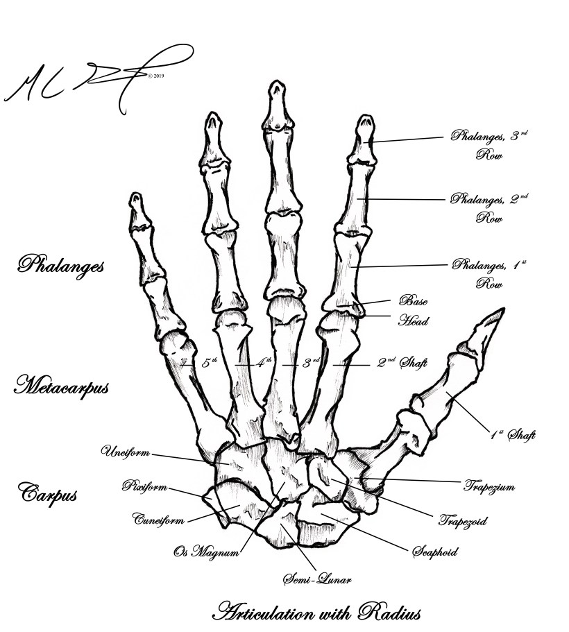
Recently deciding to branch out of my carefully carved niche of sketching fishes, I applied that approach when staring at the illustration (skeletal anatomy of the left hand by Henry Vandyke Carter, M.D.) on page 129 in my copy of “Gray’s Anatomy” by Henry Gray, FRS. Looking over the meticulously illustrated diagram can be overwhelming. And staring at it on and off over the years has not assisted in helping me learn the bones of the hand or how they articulate. So, I sketched it. I outlined my hand on a piece of paper, and started carefully filling in the bones, to the best of my ability. My proportions are likely off…and I definitely used my imagination on some parts of it. But, I sketched it. I sketched it over and over and over. My wife is surely happy that I am done with this project, because I frequently pestered her to receive her opinion (Thank you! 🙂 ). I ended up with 5 sketches in total, each with adjustments to shading in an attempt to refine my own technique and make something that I actually felt pleased with.
Left Hand, Osteology – proofs. MC Gilbert 2019
I made my best effort, and that is really all I can ask of myself. In the end, I was able to make something that I was proud of, something that I would not have been able to do three years ago. With a little photoshop magic, I was even able to tidy it up a bit and put legible labels on the bones to make a figure.
Left Hand, Osteology, Annotated. MC Gilbert 2019
Point being, this repetition and the somewhat arduous effort of creating this has helped me to understand and learn the bones of the hand and how they fit together. I hope to look back at this in three years and point out the flaws. I hope that, in three years, I am still making the effort to improve.
2019 has been unexpectedly good to me and my photography this year. I’ve made new contacts, two won awards – one from the Federation of American Societies of Experimental Biologyand the other placed in the very prestigious Images from Science 3, I sold art to a luxury condo in the Cayman Islands, and I like to think I grew as an artist. I’m fortunate to have my wife, for she is my best friend and has continually encouraged and supported me along the way (not to mention my outstanding friends and family and fans that have also supported me in numerous ways). I hope that 2020 brings just as many successes and more learning opportunities. For now, though, I have put together one last image for the year 2019. A black and white composite of three South American cichlids, two of which have occupied a great deal of my time, scientifically, over the past year and will likely continue to do so for several years to come (Images will be up for sale by the end of the day). Happy New Year!
Comparative Anatomy of Three South American Cichlids. From top to bottom – Caquetaia spectabilis, Laetacara curviceps, & Satanoperca jurupari. MC Gilbert 2019
Comparative Anatomy of Three South American Cichlids. From top to bottom – Caquetaia spectabilis, Laetacara curviceps, & Satanoperca jurupari. MC Gilbert 2019
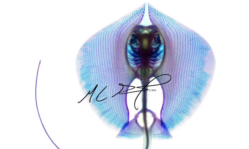
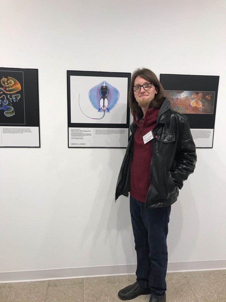
I’m beyond ecstatic to announce to everyone that my image of a cleared and stained Atlantic stingray (Dasyatis Sabina; below) was selected for entry into the very prestigious “Images from Science 3” exhibit.
I was fortunate to attend the opening of the exhibit, which took place this past weekend at the Rochester City Art Space in Rochester, New York. It was a thrilling, yet humbling experience that I was honored to share with a number of fantastic photographers, illustrators, and fellow scientists.
Standing next to my image at the RIT City Art Space in Rochester, New York on 1st Nov. 2019.
Shaking hands with Michael Peres, one of the leading organizers of “Images from Science” and Associate Chair of the School of Photographic Arts and Sciences at the Rochester Institute of Technology.
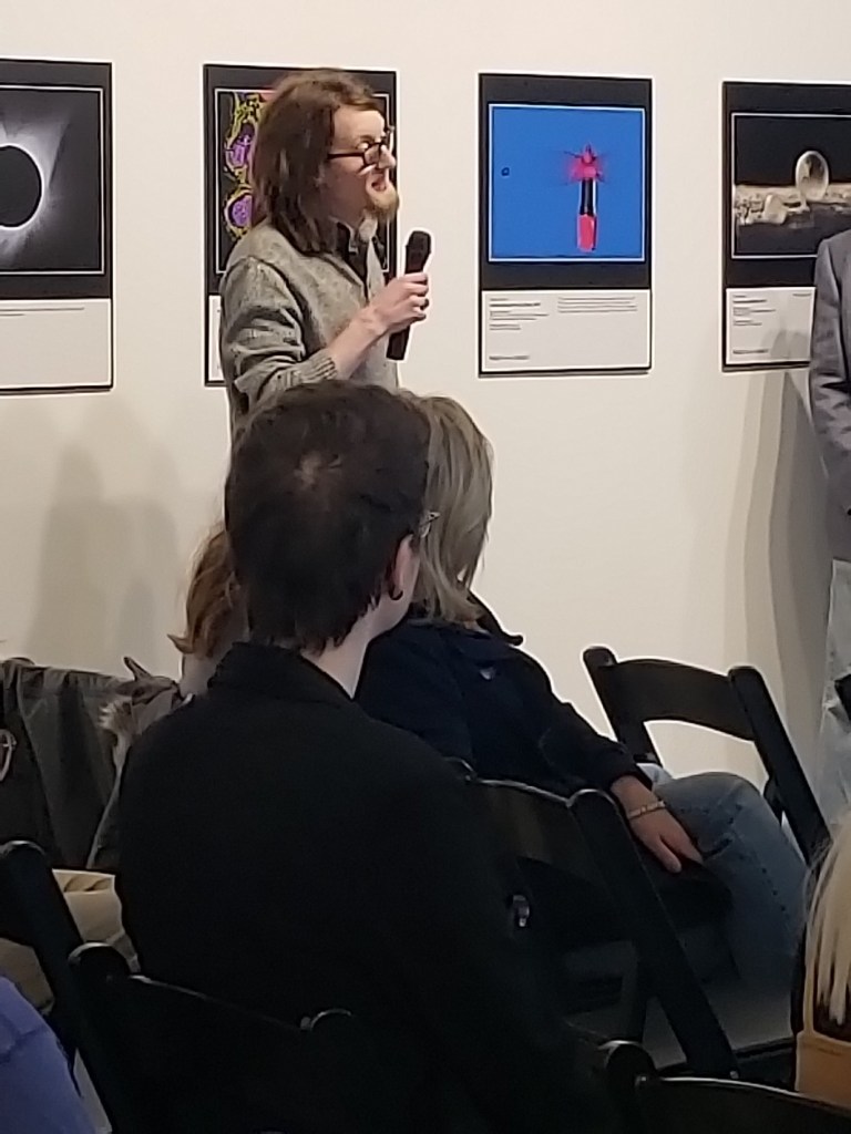
Me speaking to the exhibit visitors, organizers, and fellow contributors about my interest in photography as an evolutionary and organismic biologist.
The exhibit will remain open at the RIT City Art Space until November 24th, where it will then be moved to John Hopkins University. After an exhibit there, it will be available to travel the world for the next several years. I look forward to seeing where
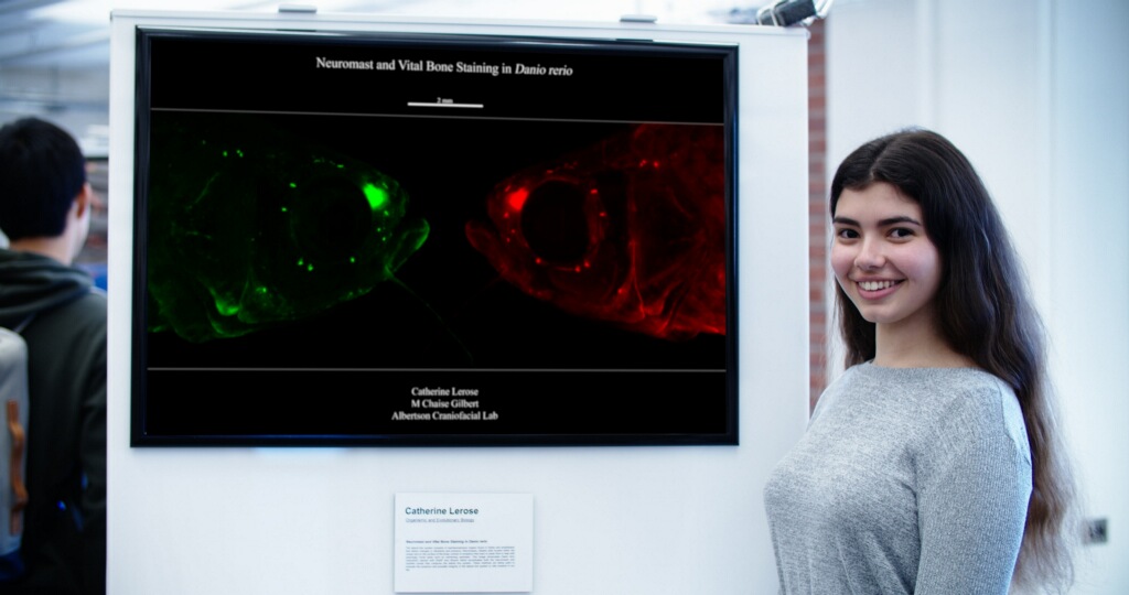
A few weeks ago, the University of Massachusetts Amherst hosted its second annual Research Art-Science Exhibition. I challenged an undergraduate working with me, Catherine Lerose, to create an artistic image that summarized what we had been working on throughout the semester. So together, we came up with a mirror image of a neuromast/bone co-stained zebrafish that we have been using to look at the lateral line canals and canal neuromasts in zebrafish. She nailed it and I am super proud of her for presenting our sciart at this event! See the full images and captions below!
Undergraduate Catherine Lerose presenting our image of a neuromast and vital bone stained zebrafish. The lateral line system consists of mechanosensory organs found in fishes and amphibians that detect changes in vibrations and pressure. Neuromasts, ciliated cells located within the canals and on the surface of the body, consist of receptors that react to water flow to help with seemingly trivial tasks such as swimming upstream. This image showcases Danio rerio (zebrafish) stained with DiASP and Alizarin which accentuates both the neuromasts and ossified canals that comprise the lateral line system. These methods are being used to evaluate the presence and possible integrity of the lateral line system in cilia mutants in our lab.
Late last year, I was surprised and honored to learn that my image of a cleared and stained zebrafish (Danio rerio) had won as a submission to the 2018 Federation of American Societies of Experimental Biology BioArt contest. There were numerous beautiful submissions to the contest and the winners were all stunning. See my full image below. Click here to see all of the phenomenal winners of the FASEB 2018 BioArt contest.
Caption from FASEB webpage:This image of a zebrafish (Danio rerio) shows the bone (green) and the cartilage (red) that comprises the skeleton. Researchers are using this image, and ones like it, to better identify how a mutation in the primary cilia can affect skeletal development, structure, and morphology.
When I first started this website, I had intended it to be little more than a hub for my photography. My images were posted to a generic page and poorly organized, my news posts were little more than ramblings and information related to sales and new images I had made, and I honestly had little to no idea what I was really wanting from a website.
Over the past two (three?) years, my abilities as a photographer and artist, as well as a scientist, have noticeably grown – if not to anyone else, then certainly to myself. I’ve taught myself new imaging techniques, I’ve picked up illustrating (albeit not quite good at it but making the effort to improve), I’ve made new contacts, I’ve began carving out a more solid niche in my own scientific community, and – perhaps most importantly – I’ve became more organized.
This website has developed in many ways since its inception back in 2016. Pages are organized, my images are neatly displayed (at least I think they are) by category and again in the order of newest in front, oldest in back, my sketches are organized by year and day of creation rather than sloppily posted in some haphazard fashion, I actually update my C.V. at least once a month, and information regarding me as a scientist can actually be found on the site now. Hell, I’ve even made a couple of figures for my own benefit that I’ve hosted here in hopes others may also benefit from them.
Point being, this site has changed a good deal, and I along with it. Something that I sincerely look forward to saying again in three years from now. I feel that it be only appropriate if I also begin using my News page for relevant … news. From presentations that I’ll be giving, to future publications, to awards when I receive them – I’ll be making my best effort to actually post information regarding me as a professional – be that as a scientist or an artist.
Thank you all for your support over the past few years and here is to another year of growing and making efforts to better improve ourselves.


You must be logged in to post a comment.